Health
Diagnosing, treating cancer with MRI: scientists

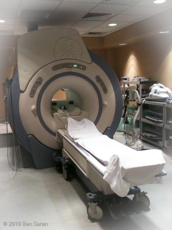
File Photo: In some cases, doctors can identify the presence of pathologies with contrast agents that locally affect the relaxation rates within damaged tissues. (Photo by Ben Saren/Flickr, CC BY-NC-ND 2.0)
MOSCOW — Researchers from the National Research Nuclear University MEPhI have recently developed a new type of contrast agents for magnetic resonance imaging (MRI) based on biodegradable silicon nanoparticles that can be used for both diagnosing and treating cancer. The results of this research were published in the Journal of Applied Physics.
An MRI is a powerful biomedical diagnostics tool based on the nuclear magnetic resonance of hydrogen atoms (protons). MRI scanners use radio waves to generate images of hydrogen atoms in the created magnetic field.
Certain methods require using contrast agents to improve the accuracy and information of the image. Contrast imaging in an MRI mostly depends on differences in the longitudinal or transverse relaxation rates.
Relaxation is the time protons need to recover to their equilibrium state. This depends on the molecules and atoms around the protons; the rate for healthy and damaged tissues varies.
In some cases, doctors can identify the presence of pathologies with contrast agents that locally affect the relaxation rates within damaged tissues.
Combining MRI and contrast agents in research helps scientists increase the possibility of imaging inflammations, such as tumorous angiogenesis in cancer.
MEPhI researchers recently developed a new type of a contrast agent based on silicon nanoparticles, which can be used for both diagnosing and treating cancer.
Viktor Timoshenko, a professor at MEPhI and Lomonosov Moscow State University, believes the new development is an example of using nanotheranostics – a combination of diagnostic and therapeutic methods applied at the nanoscale level.
Theranostic MRI contrast agents combine the effects of contrast agents and therapy conducted with medications in nanocapsules and/or additional exposure to physical fields or radiation.
“Since MRI is widely used in cancer diagnostics, developing a new type of a contrast agent, that can also be used in cancer eradication therapy, is very important for modern healthcare,” Timoshenko said.
Nanotheranostic materials must be biocompatible, not toxic to the human body. Another property is that these materials must be invisible to the immune system so that it does not destroy them right away. The nanoparticles must not accumulate in the body and their surface cannot become contaminated.
Researchers from the MEPhI Institute of Engineering Physics for Biomedicine’s Nanotheranostics Laboratory believe that using silicone nanoparticles to detect affected cells is one of the most promising methods in cancer nanotheranostics.
The nanoparticles themselves are not toxic to the human body, but, subjected to the application of radio waves, can heat up to 42оС and higher (hyperthermia), thus destroying cancer cells locally.


